Texas Plant Disease Diagnostic Lab
This is another assignment submitted by BESC484 student, B. Commer, as a partial requirement for the course. We typically get an uptick of bacterial issue showing up in the TX Plant Clinic in the fall. One of the student’s assignment is to document what they do in a manner to explain to non-plant pathologist or microbiologist what they do. Enjoy- KO
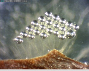
Bacterial streaming from diseased plant tissue. Bacteria was later identified as E. amylovora.
Ever wonder just how the diagnostician can confirm bacterial plant pathogens? It starts with a very simple yet fascinating test to check for what is called “streaming.” Symptomatic samples are thoroughly examined under the microscope for abrupt color changes signifying a transition from infected to healthy tissue. Once the transition zones are located, a sterile razor blade is used to cut a wedge into the vascular tissue of the stem or directly through a necrotic leaf spot. These pieces are then transferred to a droplet of sterile distilled water vascular side down, usually in a petri dish, and observed again under the dissecting microscope. Over the course of five to ten minutes (Editorial correction – on severely infected tissue – streaming would occur within 5-10 seconds of contact with water), bacterial populations will be observed exiting, or streaming, from infected areas of tissue. This can easily be confused with plant material, which also exits the fresh wound, but the bacteria will usually have a slight color tint to differentiate it. Tiny bubbles can also be associated with the streaming and should be watched closely for accompanying bacteria. If streaming is noticed, a sterile loop is swirled into the water droplet and streaked onto nutrient agar media plates (one plate per positive droplet). After roughly one day in a bacterial incubator, single isolated colonies should be transferred to TSBA plates, also one colony per new plate. It is a good idea at this stage to choose colonies with differing characteristics or colors so that the pathogenic variety is not overlooked in the purification process. 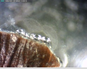 After a strict 24 hour time period (give or take one hour), the cultures are harvested and placed in sterile vials for identification using a Microbial Identification System to perform fatty acid methyl ester analysis by gas chromatograph and match the bacteria to those stored in a mass library index. The following pictures are streaming from a Bradford Pear twig infected with Erwinia amylovora, the causal agent of many vascular wilts. To learn more about the disease and pathogen, check out our Fire Blight factsheet:
http://plantclinic.tamu.edu/factsheets/fireblight/